Radial tuberosity
| Radial tuberosity | |
|---|---|
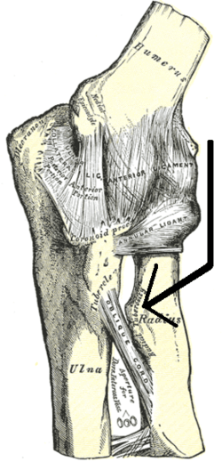 Left elbow-joint, showing anterior and ulnar collateral ligaments. (Radial tuberosity visible at center right.) | |
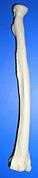
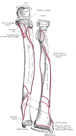 Bones of left forearm. Anterior aspect. (Radius is bone on right. Radial tuberosity is visible at upper left of radius.) | |
| Details | |
| Identifiers | |
| Latin | Tuberositas radii |
| TA | A02.4.05.007 |
| FMA | 23489 |
Beneath the neck of the radius, on the medial side, is an eminence, the radial tuberosity; its surface is divided into:
- an anterior, rough portion, for the insertion of the tendon of the biceps brachii.
- a posterior, smooth portion, on which a bursa is interposed between the tendon and the bone.
Sources
This article incorporates text in the public domain from the 20th edition of Gray's Anatomy (1918)
External links
- Anatomy figure: 07:02-08 at Human Anatomy Online, SUNY Downstate Medical Center
- radiographsul at The Anatomy Lesson by Wesley Norman (Georgetown University) (xrayelbow)
Additional images
 Radial tuberosity shown.
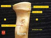 Anterior View. Radial tuberosity.
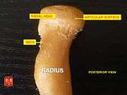 Posterior View. Radial tuberosity.
 Radial tuberosity below neck.
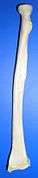 Radial tuberosity below neck.
 Left Radius - close-up - animation.
.jpg) Elbow joint - deep dissection (anterior view, human cadaver)
Elbow joint - deep dissection (anterior view, human cadaver)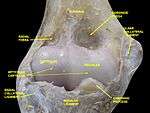 Elbow joint - deep dissection (anterior view, human cadaver)
This article is issued from Wikipedia - version of the 9/13/2015. The text is available under the Creative Commons Attribution/Share Alike but additional terms may apply for the media files.